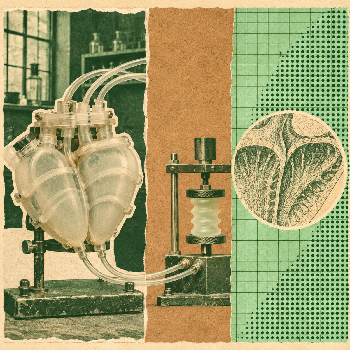

Researchers at UNSW Sydney have developed a fully synthetic, soft robotic human heart that replicates the organ’s complex movements and internal structures. Presented as a realistic environment for testing the next generation of cardiac devices, this beating model is the subject of two publications, in Nature Communications and in Advanced Science.
The model replicates the left side of the heart, with artificial valves, papillary muscles, and tendon cords (chordae tendineae), structures essential to the organ’s proper function and often affected by disease. According to the researchers, it can reproduce what happens when valves leak and blood flows backward, a phenomenon that increases the risk of heart failure and other potentially fatal complications. Team leader Thanh Nho Do, Scientia Associate Professor at UNSW’s School of Biomedical Engineering and a member of the UNSW Medical Robotics Lab, notes that cardiovascular diseases remain the leading cause of death worldwide.
A Soft, Beating Heart
The device is a soft, flexible replica of the left side of the heart. Silicone membranes form the internal cavities, while soft robotic artificial muscles, coiled around the structure, reproduce the way the heart contracts and twists. These muscle fibers are powered by hydraulic pressure, which the researchers control to move the model. According to Dr. James Davies, a postdoctoral fellow in Thanh Nho Do’s group, “the model is made from soft materials and actuated by artificial muscles arranged to mimic the layered muscle architecture of the human heart.”
Unlike conventional laboratory models, this heart incorporates the structures that control the mitral valve, which opens and closes with each beat to circulate oxygen-rich blood throughout the body while preventing backflow. The system also allows adjustment of the tension in the artificial papillary muscles that support this valve. The teams were thus able to recreate conditions close to disease, such as mitral valve prolapse and regurgitation, where blood flows backward instead of circulating properly.
Measurements Close to Human
Using ultrasound and measurements of blood pressure and flow, the researchers indicate that the model behaves very similarly to a human heart. A healthy valve produces normal pressure and flow profiles, while induced disease leads to characteristic changes observed in patients. The model was also tested with a recently developed soft cardiac catheter, which was able to navigate inside the artificial heart and detect contact with moving structures.
For the researchers, the platform could reduce reliance on animal studies, at least in the early phases of medical device development. Thanh Nho Do believes it would allow “replicating specific heart diseases, such as heart failure with preserved ejection fraction (HFpEF), which remains one of the least understood and most difficult to treat.” The model reproduced several changes observed in this disease, including a reduced ability of the heart to relax between beats, which slows filling and increases internal pressure.
In the longer term, the researchers envision personalized versions of the model, built from medical imaging data, to help clinicians compare devices and treatments before surgery.
Validation Still Needed
The researchers emphasize, however, that the model remains a proof of concept and not a finished clinical tool. Materials, control systems, and compatibility with medical imaging need improvement, as does the use of patient-specific geometries. Thanh Nho Do believes the next essential step is thorough validation against clinical data.
The team, which also includes Professor Christopher Hayward, a cardiologist specializing in heart failure and transplantation, as well as Professors Jelena Rnjak-Kovacina and Hoang-Phuong Phan, hopes the technology can be adopted in clinical settings. “Rather than considering the current model as a finished clinical tool, we see it as an enabling platform,” concludes James Davies.
